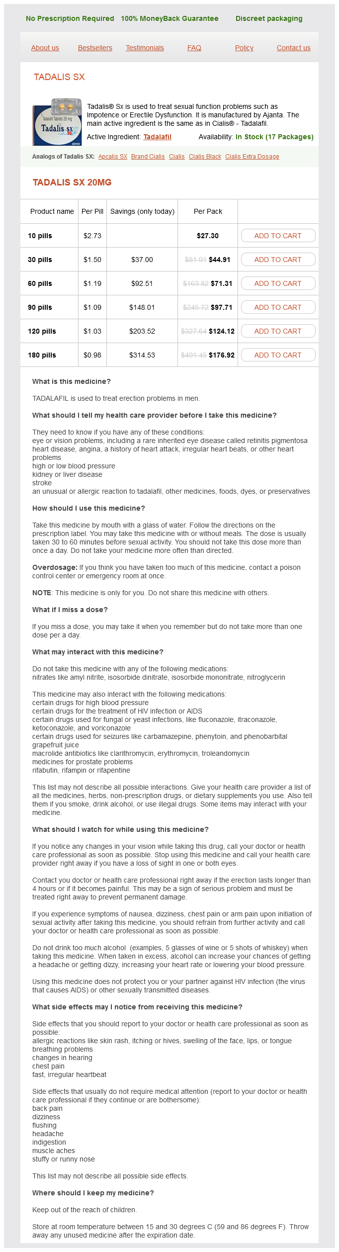
Tadalis SX 20mg
- 10 pills - $27.30
- 30 pills - $44.91
- 60 pills - $71.31
- 90 pills - $97.71
- 120 pills - $124.12
- 180 pills - $176.92
Tadalis SX dosages: 20 mg
Tadalis SX packs: 10 pills, 30 pills, 60 pills, 90 pills, 120 pills, 180 pills
Only $1.18 per item
In stock: 641
Description
It is the most sensitive test for both congenital and acquired colour vision defects erectile dysfunction caused by ptsd trusted tadalis sx 20 mg. It consists of 85 hue caps (not 100) and colour vision is judged by error score (higher score means poorer colour vision). Farnsworth D15 hue discrimination test is similar test but utilizes only 15 hue caps. It is also a spectroscopic test where a central coloured plate is to Chapter 13 Neuro-ophthalmology 331 be matched to its closest hue from four surrounding colour in each of 10 plates. In this test, the observer is asked to mix red and green colour in such a proportion that the mixture should match the given yellow coloured disc. The judgement about the defect is made from the relative amount of red and green colours and the brightness setting used by the observer. In this, the subject is asked to make a series of colour-matches from a selection of skeins of coloured wools. AmAurosis It implies complete loss of sight in one or both eyes, in the absence of ophthalmoscopic or other marked objective signs. AmblyoPiA It implies a partial loss of sight in one or both eyes, in the absence of ophthalmoscopic or other marked objective signs. It may be anisometropic, strabismic or due to stimulus deprivation (amblyopia ex anopsia) (see page 342). Causes of cortical blindness include: It refers to a sudden, temporary and painless monocular visual loss occurring due to a transient failure of retinal circulation. An attack of amaurosis fugax is typically described by the patients as a curtain that descends from above or ascends from below to occupy the upper or lower halves of their visual fields. When observed shortly after an attack, the fundus may either be normal or reveal signs of retinal ischaemia such as retinal oedema and small superficial haemorrhages. Cortical blindness is characterized by: · Bilateral loss of vision, · Normal pupillary light reflexes, · Visual imagination and visual imagery in dream are preserved, · Anton syndrome, i. It is a sudden, bilateral, complete loss of sight occurring probably due to the effect of certain toxic materials upon the cells of the visual centre in patients suffering from acute nephritis, eclampsia of pregnancy and renal failure. The fundi are usually normal except for the coincidental findings of hypertensive retinopathy, mAlingering In malingering a person poses to be visually defective, while he is not. Usually, one eye is said to be blind which does not 332 Section iii Diseases of Eye the symptoms and negotiates well with the surroundings (c. Differential diagnosis Before diagnosing malingering following conditions (which produce visual loss with apparently normal anterior segment and a normal fundus) should be ruled out: 1. Cortical blindness must be ruled out from its characteristic features (see page 331). Chiasmal tumours may sometimes present with visual loss and normal fundus (before the onset of optic atrophy).
Syndromes
- Drowning
- Loss of social engagement
- Treating allergies by staying away from triggers (such as dust). Older children may be given allergy medications.
- Muscle weakness
- MRI of the neck
- Post-pericardiotomy syndrome (low-grade fever and chest pain) that could last up to 6 months
- Potassium hydroxide
- Shortness of breath that gets worse with activity
Most of these patients also have ventricular septal defects erectile dysfunction treatment hypnosis tadalis sx 20 mg line, pulmonary valve abnormalities, and/or Ebsteinoid changes of the tricuspid valve. The traditional surgical approach ("functional repair") has been to repair the associated lesions only. This leaves the patient with a morphologic right ventricle and tricuspid valve as the systemic ventricle and atrioventricular valve. More recently, some centers have advocated an anatomic repair, the "double switch" procedure, in certain subgroups of these patients. Patients with two adequate ventricles and a normal pulmonic valve undergo an arterial switch procedure combined with a Senning or Mustard atrial switch. If the pulmonic valve is not suitable for an arterial switch, a Senning and Rastelli procedure may be an option if there is an appropriate ventricular septal defect for baffling the morphologic left ventricle to the aortic valve. Theoretically, a double switch procedure should improve the long-term outcome of these patients, who often develop progressive tricuspid regurgitation and right ventricular failure, by making the morphologic left ventricle the systemic ventricle and placing the abnormal tricuspid valve in the lower pressure pulmonary circulation. However, proper patient selection is critical, and many patients require a multistaged pulmonary banding procedure to train the left ventricle. When performing the double switch operation, either the arterial switch or the atrial switch can be performed first. Some modifications to both procedures may be required because of previous pulmonary artery banding and anatomic considerations. Closure of Ventricular Septal Defect the ventricular septal defect is closed through the mitral valve. Sutures are placed on the morphologic right ventricular side of the septum to avoid the conduction system. Previous Pulmonary Banding Dissection between the aorta and pulmonary artery should be carried out carefully in the presence of a previously placed pulmonary band. This may require Vshaped excisions of tissue from the sinuses before coronary transfer to ensure a competent valve. In addition, the area of the band must be excised or enlarged to prevent supravalvar stenosis and distortion of the sinotubular junction. The site of the superior vena cava cannula should be as high as possible above the cavoatrial junction. Individual caval cannulation must be done carefully to minimize interference with venous return and avoid hypotension and serious dysrhythmias. Partial cardiopulmonary bypass may be initiated after one cannula is inserted to facilitate placement of the second cannula. Under moderate hypothermia, the aorta is cross-clamped and cardioplegia is delivered into the aortic root. Atrial Incision Access to the inside of the right atrium is gained through a longitudinal incision made 3 to 4 mm anterior and parallel to the sulcus terminalis.
Specifications/Details
This is accomplished with interrupted 3-0 Ticron sutures buttressed with Dacron or pericardial pledgets on both the atrial and ventricular sides of the sutures vegetable causes erectile dysfunction 20 mg tadalis sx buy amex. Right Coronary Artery Injury the ventricular plication sutures must be placed carefully after identifying the right coronary artery and its branches to avoid direct injury to , or distortion of, the coronary arteries, which can result in myocardial infarction. Injury to Conduction Tissue Because of the proximity of the atrioventricular node to the bundle of His, placement of sutures that extend between the septal leaflet and the true right atrium is hazardous, particularly to the left of the coronary sinus. Creation of an Aneurysmal Cavity the mattress sutures are woven in and out of the atrialized portion of the right ventricle so that when they are tied, the atrialized ventricle is completely obliterated and no aneurysmal chamber is formed. Bicuspidization Depending on the anatomy, it is sometimes possible to exclude the posterior leaflet by a modified annuloplasty converting the tricuspid valve into a bicuspid valve (or if the septal leaflet is very dysplastic, a monocuspid valve), thereby eliminating any residual tricuspid insufficiency. This is achieved by constricting the posterior segment of the annulus with interrupted sutures of 3-0 Ticron buttressed with pledgets. An alternate technique championed by Carpentier entails temporary detachment of the anterior leaflet from the anteroseptal commissure to the junction with the posterior leaflet, if present. Extensive mobilization is achieved by dividing the fibrous band connections of the leaflets to the muscular wall of the right ventricle. It is plicated with multiple interrupted sutures of 3-0 Ticron, and the posterior annulus is similarly reduced in size to achieve a relatively normal right ventricular geometry. The redundant atrial wall behind the coronary sinus may also need to be plicated with continuous suture of 4-0 Prolene. The anterior leaflet is then reattached to the fibrous annulus using continuous 5-0 Prolene suture. This repair is often reinforced with an annuloplasty ring after clockwise rotation of the valve that uses the redundant anterior leaflet to augment the deficiency along the septal leaflet. Excess Traction on the Leaflets If the anterior papillary muscle is malpositioned, it must be cut at its base and reimplanted at a higher level in the septum or the ventricular wall with pledgeted 3-0 Prolene sutures. Injury to the Conduction System the right atrial plication must be accomplished to the right side of the coronary sinus to avoid the conduction system. Bidirectional Cavopulmonary Anastomosis If right ventricular function is impaired, a bidirectional cavopulmonary anastomosis can decrease the right ventricular preload and may improve patient survival. This procedure does eliminate catheter access from the upper extremities in these patients who may need future ablation procedures or pacemakers. Because some of these patients have associated left ventricular dysfunction, it is important to document low left atrial and pulmonary pressures before performing a bidirectional cavopulmonary shunt (see Chapter 31). Atrial Septal Defect Closure the patent foramen ovale or atrial septal defect should be closed by primary suture or patching with pericardium or Gore-Tex (see Chapter 19). After standard bicaval cannulation and cardioplegic arrest, the anatomy is inspected. The anterior leaflet is then taken down, in particular, capitalizing on the true area of delamination between the anterior leaflet and the right ventricle. All fibrous and muscular attachments of the leading edge of the leaflet are extensively taken down, with attention paid not to divide true chordal support to the leading edge. The dissection is then extended clockwise to include the septal leaflet all the way to the anteroseptal commissure; the septal leaflet often is quite diminutive (nearly absent) and may have multiple fenestrations that require closure.
Sarracenia purpurea (Pitcher Plant). Tadalis SX.
- Are there safety concerns?
- Dosing considerations for Pitcher Plant.
- How does Pitcher Plant work?
- What is Pitcher Plant?
- Digestive disorders, constipation, urinary tract diseases, fluid retention, preventing scar formation, pain, and other conditions.
Source: http://www.rxlist.com/script/main/art.asp?articlekey=96145
Related Products
Usage: q.h.
Additional information:
Tags: cheap tadalis sx 20 mg mastercard, 20 mg tadalis sx order mastercard, cheap 20 mg tadalis sx fast delivery, generic 20 mg tadalis sx amex
10 of 10
Votes: 313 votes
Total customer reviews: 313
Customer Reviews
Roy, 39 years: However, it is often advisable to create an opening on the aortic side of the graft matching the aortic opening and to perform a side-to-side anastomosis. Without a doubt, stimulus delivery to the site of interest will profoundly affect the overall energy requirements. If too little graft remains, the suture line may distort or compromise flow into the graft from the aorta. If the patient passes this, they are then recommended to undertake further testing, including an on-the-road test, at a regional driving centre to ensure road safety and to be assessed and advised on any physical adaptations required to their vehicle.
Kan, 60 years: If a shunt procedure is not performed, the ductus arteriosus can be left open and only temporarily occluded during cardiopulmonary bypass. The syntrophin mutation may disrupt this complex and lead to increased nitrosylation of the channel, thus increasing the Nav1. Extraocular dislocation may be in the subconjunctival space (phacocele) or it may fall outside the eye. Prevention of Heart Block the bundle of His pierces the central fibrous body and the tricuspid annulus before crossing into the ventricular septum and following a course along the inferior margin of the defect toward the left ventricular side of the septum.
Umbrak, 61 years: Other congenital defects can also be associated with transposition of the great arteries. Occasionally, the leaflets are thickened and dysplastic and produce obstruction by their bulkiness. Additional reviews on the characteristics of these structures can be found elsewhere. In Jänig W, editor: the Integrative Action of the Autonomic Nervous System: Neurobiology of Homeostasis, Cambridge, 2006, Cambridge University Press, pp 1334.
Lisk, 63 years: Of note, preganglionic neurons may synapse on neurons within the ganglia at the same thoracic level or may travel within the sympathetic chain and synapse on neurons of ganglia at other spinal levels. The anterior leaflet is then taken down, in particular, capitalizing on the true area of delamination between the anterior leaflet and the right ventricle. It affects individuals of all ages with higher incidence in the older individuals, which is in part due to idiopathic degenerative disease. Recent genome-wide association studies have, however, found strong associations with a couple of gene loci, suggesting that genetic factors do play a part.
Spike, 59 years: Orofacial abnormalities include flattened nasal bridge, maxillary hypoplasia, cleft palate and high arched palate. The aortic valve may lie quite close below the valve, and with the aorta decompressed may fall closer to the tricuspid valve, making it more likely to be subject to injury. It consists of dilated vascular channels and does not grow or regress like the capillary haemangioma. Li D, Zhang L, Kneller J, et al: Potential ionic mechanism for repolarization differences between canine right and left atrium.
Nerusul, 50 years: Intravitreal drug implants are inserted in the vitreous cavity through pars plana for sustained and slow release. Practically, this is achieved by increasing the number of ultrasound pulses emitted. Indications: It is a very good alternative to pilocarpine in resistant or intolerant cases. These symptoms occur either due to acute secondary glaucoma or apparent intraocular inflammation or orbital cellulitis.
Temmy, 56 years: Three most common forms of nystagmus seen in childhood begin in infancy and therefore, are not congenital as believed earlier. These lenses lie entirely in front of the iris and are supported in the angle of anterior chamber. These electrodes can be displayed simultaneously in isolation or relative to the reconstructed 3D chambers. However, if the number of fibroblasts was locally increased or the number of cardiomyocytes was locally decreased, such as in fibrotic disease, this protective balance would be compromised and the fibroblast paracrine action would negatively affect the electrical function of the surrounding cardiomyocytes and potentially lead arrhythmic activity.
Dan, 58 years: If this is done through the posterior (pulmonary) valve, great care must be taken to avoid the conduction system. Kehat I, Kenyagin-Karsenti D, Snir M, et al: Human embryonic stem cells can differentiate into myocytes with structural and functional properties of cardiomyocytes. Second, by roving the mapping catheters, 3D renderings of various cardiac chambers can be created. Causes of bilateral ptosis include congenital ptosis, myasthenia gravis, myotonic dystrophy, KearnsSayre syndrome, Lambert-Eaton myasthenic syndrome, and chronic progressive external ophthalmoplegia.