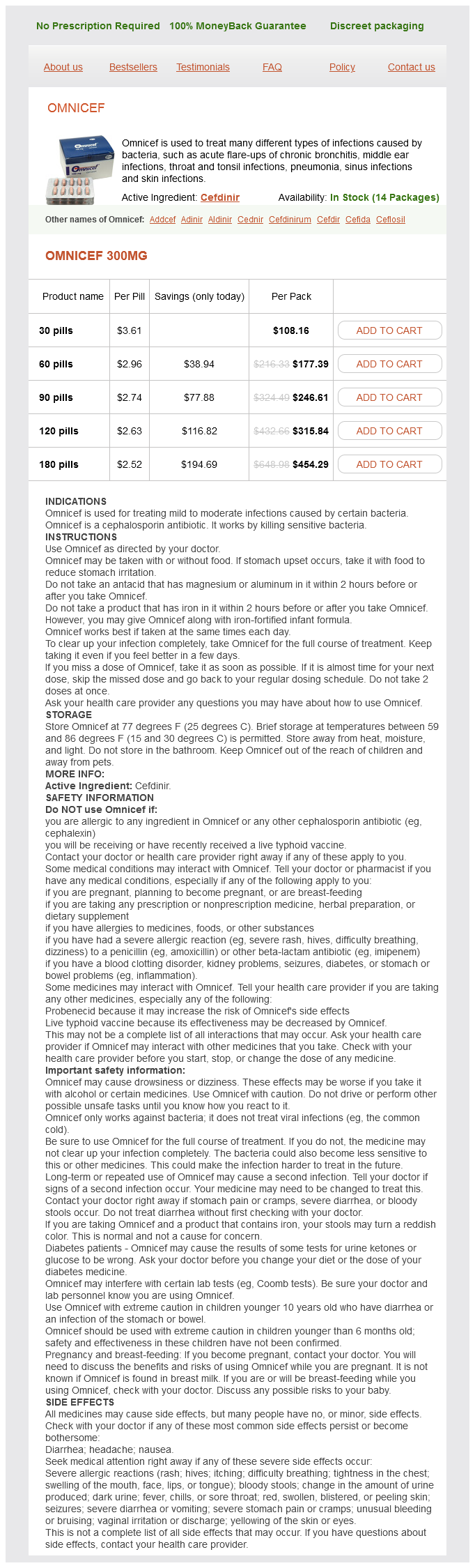
Omnicef 300mg
- 30 pills - $108.16
- 60 pills - $177.39
- 90 pills - $246.61
- 120 pills - $315.84
- 180 pills - $454.29
Omnicef dosages: 300 mg
Omnicef packs: 30 pills, 60 pills, 90 pills, 120 pills, 180 pills
Only $2.68 per item
In stock: 757
Description
Palpating lateral to flexor carpi radialis antibiotic qt prolongation discount 300 mg omnicef with mastercard, 34 cm proximal to the wrist crease, reveals the muscle belly of flexor pollicis longus (flexing and extending the thumb will confirm that the examining finger is correctly placed). The area on the ulnar side of flexor carpi radialis tendon is packed with functionally important structures. The median nerve is either covered by, or situated just lateral (radial) to , the tendon of pal maris longus. Near to the wrist, the median nerve lies very close to the skin and can, there fore, be injured by relatively superficial lacerations. When palmaris longus is absent, only a thin covering of subcutaneous fat and deep fascia separate skin and nerve. The four tendons of flexor digitorum superficialis lie deep to the median nerve; the tendons to the middle and ring fingers lie anterior to those for the index and little fingers as they pass deep to the flexor retinaculum, and can be felt and, usually, seen to move during finger flexion/extension. The snuffbox is bounded on its anterolateral (radial) side by the tendons of abductor pollicis longus (laterally) and extensor pollicis brevis (immediately medial), and on its posteromedial/ulnar side by the tendon of extensor pollicis longus. Running a finger along extensor pollicis brevis enables palpa tion of the superficial radial nerve, which can be rolled from side to side on the tendon. The cephalic vein passes over the roof of the snuff box, where it is visible and palpable, and the pulsation of the radial artery can be palpated deeply on its floor. Compartments 5 (containing extensor digiti minimi) and 6 (containing extensor carpi ulnaris) flank the posterior and medial sides of the ulnar head, respectively. Compartment 4 (containing exten sors digitorum and indicis) sits between the ulnar head and compart ment 3. The tendon of extensor carpi ulnaris is palpable distal to the ulnar styloid as it crosses the wrist, with the wrist in extension and adduction. The first dorsal interosseous forms a visible fleshy mass on the lateral part of the dorsal hand between the index finger and thumb, especially whilst abducting the index finger against resistance. Its pulsations are palpable along this line of travel and proximal to the bicipital aponeurosis, via com pression against the humerus. Brachial artery catheterization just proxi mal to the aponeurosis is used for coronary angiography or cardiac catheterization. The ability to compress the brachial artery against the humerus makes the arm the favoured site for noninvasive blood pres sure measurement. The median nerve is intimately related to the bra chial artery throughout its course in the arm. Proximally, it sits lateral to the artery; then, around the midpoint of the arm, it passes anterior to the artery and descends on its medial side to the cubital fossa. From here, the ulnar artery passes inferomedially to a point onethird of the way down flexor carpi ulnaris (which passes along a line joining the medial humeral epi condyle to the pisiform). The ulnar artery and nerve then pass inferiorly to a point just lateral to the pisiform along the line of flexor carpi ulnaris, with the ulnar artery lying on the lateral (radial) side of the nerve. The radial artery passes inferolaterally through the forearm, from its point of origin to a point just lateral to the tendon of flexor carpi radialis, anterior to the distal radius. The upper part of its course can be represented by a line that passes deep to the medial part of the eleva tion produced by brachioradialis on the anterior aspect of the forearm.
Syndromes
- Have you had any open sores?
- Is it getting worse or more noticeable?
- Loses temper
- Decreased ability to fight infection
- Poor gag reflex in people who are not alert (unconscious or semi-conscious) after a stroke or brain injury
- Irregular menstrual bleeding or spotting
- Bone pain and fever
- For a total or simple mastectomy, the surgeon cuts breast tissue free from the skin and muscle and removes it. The nipple and the areola are also removed. The surgeon may do a biopsy of lymph nodes in the underarm area to see if the cancer has spread. In some rare breast cancers, a simple mastectomy is performed on both breasts.
Laterally antibiotic bronchitis omnicef 300 mg order with mastercard, it lies under longissimus capitis and sometimes splenius capitis, both of which overlap obliquus capitis superior. The vertebral artery and the dorsal ramus of the first cervical nerve lie in a groove on the upper surface of the posterior arch of the atlas. VascularsupplyThe suboccipital muscles receive their blood supply from the vertebral artery and deep descending branches of the occipital artery. They lie inferior to the anterior part of the occipital bone, where three of the muscles form the boundaries of the suboccipital triangle: rectus capitis posterior major lies above and medially; obliquus capitis superior lies above and laterally, and obliquus capitis inferior lies below and laterally. With the head in the anatomical position, the suboccipital triangle lies almost in the horizontal plane. Axial rotation involves twisting of vertebrae relative to each other with accompanying torsional deformation of intervening discs. Elsewhere in the column, although movement is slight between individual vertebrae, the range summates to become considerable for the column as a whole. In the postcervical column, the effective range of rotation is greatest at the thoracolumbar junction. In the cervical region, the upward inclination of the superior articular facets allows free flexion and extension. The latter is usually greater, and is checked above by locking of the posterior edges of the superior facets of C1 in the occipital condylar fossae, and below by slipping of the inferior processes of C7 into grooves inferoposterior to the first thoracic superior articular processes. Flexion stops where the cervical convexity is straightened, checked by apposition of the projecting lower lips of vertebral bodies on subjacent bodies. Cervical lateral flexion and rotation are always coupled, and the superomedial inclination of the superior articular facets imparts rotation during lateral flexion. Two physiological movements take place at the atlantooccipital joints: those of flexionextension and lateral flexion. Some studies have suggested that maximum flexionextension occurs between the occiput and C1; however, Frobin et al (2002) noted between 12. Global cervical flexion ranges from 45° to 58°, depending on the method of assessment, age and sex; older subjects and females exhibit less motion (Ordway et al 1997, Trott et al 1996). At an intersegmental level, motion increases from the second cervical level and peaks at the mid-cervical level (1417° recorded at C4/5), before reducing at the junction of the cervical and thoracic spine (9. Global ranges of lateral flexion range from 32° to 47°, with a gradual reduction in range with age and sex, while rotational movements range from 63° to 78°. In the thoracic region, especially superiorly, all movements are limited, reducing interference with respiration. Lack of upward inclination of the superior articular facets prohibits much flexion, and extension is checked by contact of the inferior articular margins with the laminae and of adjacent spines. Its axis is in the vertebral bodies in the mid-thoracic region, and in front of them elsewhere, so that rotation involves some lateral displacement. The direction of articular facets would allow free lateral flexion but this is limited in the upper thoracic region by the resistance of the ribs and sternum. Movement in the thoracic spine is frequently regionalized to upper, mid- and lower thoracic.
Specifications/Details
It stretches back on either side from the midlevel of the thyroid angle to the vocal processes of the arytenoids infection ear piercing 300 mg omnicef free shipping. The vocal folds lie on either side of a fissure, the rima glottidis, and form the anterolateral three-fifths of its edges. The posterior two-fifths of the edges of the rima glottidis are formed by the vocal processes of the arytenoid cartilages (to which the vocal folds are attached). The mucosa overlying the vocal ligament is thin and attached to the underlying lamina propria by a basement membrane. It lies directly on the ligament, and so the vocal fold appears pearly white in vivo. It becomes a bilaminar structure by 2 months of age and three layers become established by 7 years of age (Hartnick 2005). The intermediate layer consists of elastic fibres, and the deep layer is formed of collagen fibres; these two layers collectively form the vocal ligament. The appearance of differential fibres namely, elastin and collagen is noted at 13 years of age (Hartnick 2005). Fibres of thyroarytenoid and vocalis form the fifth layer of the vocal folds; they shorten, relax and aid adduction of the vocal folds (see p. The site where the vocal folds meet anteriorly, the anterior commissure, is the region where fibres of the vocal ligament pass through the thyroid cartilage to blend with the overlying perichondrium. The point at which the vocal ligaments attach to the thyroid cartilage is known as Broyles ligament; it contains blood vessels and lymphatics, and therefore represents a potential route for the escape of malignant tumours from the larynx. This is a very significant anatomical escape pathway for primary tumours arising on the vocal cord. Located at the anterior and posterior end of each vocal ligament are the maculae flavae. These form conspicuous mucosal bulges visible on endoscopic examination of the larynx through the mucosa as whitish yellow masses. The vocal folds are connected to thyroid cartilage anteriorly via the anterior maculae flavae and the anterior commissure tendon, and posteriorly via the posterior maculae flavae. The maculae flavae themselves are described as being formed of dense masses of stellate cells with a morphology markedly different from that of fibroblasts surrounded by a dense extracellular matrix. The function of the maculae flavae remains unclear but it has been suggested that they play a critical role in the growth, development and metabolism of the extracellular matrix of the vocal folds (Awd Allah et al 2009, Fayoux et al 2004, Sato et al 2010a, 2010b). Laryngeal vestibule the laryngeal vestibule is the region between the laryngeal inlet and vestibular folds. The anterior wall is formed by the posterior surface of the epiglottis, the lower part of which (epiglottic tubercle) bulges backwards a little. The lateral walls, higher in front and shallow behind, are formed by the medial surfaces of the aryepiglottic folds. The posterior wall consists of the interarytenoid mucosa above the ventricular folds. On each side it contains the vestibular folds, the ventricle and the saccule of the larynx.
Dahlia Inulin (Inulin). Omnicef.
- What is Inulin?
- Lowering high levels of a kind of fat called triglycerides.
- Dosing considerations for Inulin.
- How does Inulin work?
- Are there safety concerns?
- Weight loss.
- Constipation.
- What other names is Inulin known by?
- High cholesterol levels.
Source: http://www.rxlist.com/script/main/art.asp?articlekey=97001
Related Products
Usage: p.r.n.
Additional information:
Tags: omnicef 300 mg buy low cost, buy omnicef 300 mg visa, order 300 mg omnicef free shipping, buy generic omnicef 300 mg
10 of 10
Votes: 215 votes
Total customer reviews: 215
Customer Reviews
Ashton, 62 years: The posterior auricular artery ascends between the parotid gland and the styloid process to the groove between the auricular cartilage and mastoid process. The canalis sinuosus descends in the orbital floor lateral to the infraorbital canal, curves medially in the anterior wall of the maxillary sinus, and then passes below the infraorbital foramen to the margin of the anterior nasal aperture in front of the anterior end of the inferior concha. Actions Acting from below, scalenus medius bends the cervical part of the vertebral column to the same side.
Kippler, 32 years: The posterior axillary fold, produced by latissimus dorsi and the underlying teres major, reaches a lower level on the humerus than the anterior axillary fold. Orbital connective tissue pulleys There is mounting evidence that challenges the traditional view that the recti are attached only at their origin and scleral insertion. The lesser petrosal nerve, which may be regarded as the continuation of the tympanic branch of the glossopharyngeal nerve, traverses the tympanic plexus.
Emet, 45 years: The vascular mesenchyme, which enters the cup through the choroidal fissure and around the equator of the lens, associates locally with this reticular tissue and thus contributes to the formation of the vitreous body. Key: 1, superior articular facet of S1; 2, ala; 3, first dorsal sacral foramen; 4, attachments of interosseous sacroiliac ligaments; 5, lateral crest and transverse tubercles; 6, median crest and spinous processes; 7, attachment of gluteus maximus; 8, sacral hiatus; 9, cornua; 10, intermediate crest and articular tubercle (inferior articular process); 11, posterior surface of body of S1 forming anterior wall of sacral canal; 12, area of attachment of multifidus (bounded by thin line); 13, attachment of erector spinae aponeurosis (thick line). Neonatal extraocular muscle coordination is usually achieved by 36 months of age and persistent deviation of an eye requires evaluation.
Ivan, 21 years: Lacrimal drainage pathway There is a constant turnover of tears; production is matched by elimination. Tonsillar involution begins at puberty, when the reactive lym phoid tissue begins to atrophy, and by old age only a little tonsillar lymphoid tissue remains. The third part of the subclavian artery descends laterally from the lateral margin of scalenus anterior to the outer border of the first rib, where it becomes the axillary artery.
Grimboll, 60 years: In the thoracic region they are nearly uniform, and the anterior concavity is largely due to the vertebral bodies. A third often ascends medial to the vertebral artery in front of the seventh cervical transverse process. It has been suggested that vascular loops in the internal acoustic meatus (from the anterior inferior cerebellar artery) might generate pulsatile tinnitus.
Thorek, 37 years: LigamentsLigaments that work in conjunction with, and modify the function of, the facet joints throughout the vertebral column are described on pages 731733. The fundamental frequency and its associated harmonics may also be raised or lowered by appropriate elevation or depression, respectively, of the hyoid bone and the larynx as a unit by the selective actions of the extrinsic laryngeal muscles. Vascular supply the arterial supply of the superior constrictor is derived mainly from the pharyngeal branch of the ascending pharyn geal artery and the tonsillar branch of the facial artery.
Pranck, 24 years: The latter serves as a useful landmark for the inferior limit of the adult dural sac. The medial plate is functionally related to the pharynx, providing attachment for the pharyngobasilar fascia, superior constrictor and pterygomandibular raphe. When the eye is open, the tear film is in a state of equilibrium with the oxygen in the atmosphere, and gaseous exchange takes place across the tearepithelial interface.
Milten, 53 years: The nasopharynx is a narrow tube that curves gradually to join the oropharynx without any sharp junctional demarcation. It ascends between their transverse processes, passes anterior to the first posterior intertransverse muscle and emerges lateral to the vertebral artery, generally between longus capitis and levator scapulae. The nerve may be damaged during parotid gland surgery, resulting in impaired sensation of the tragus and temple.