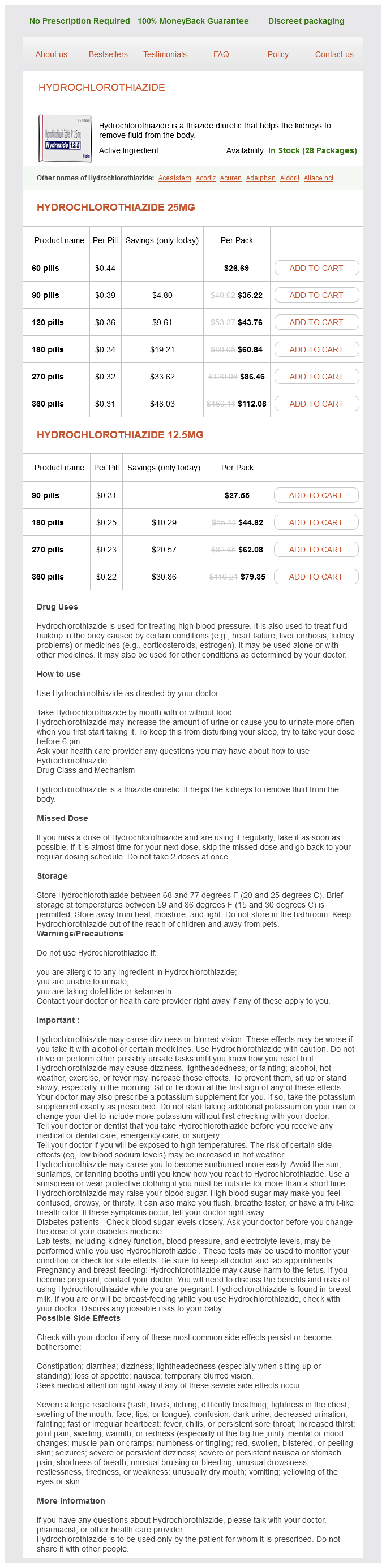
Hydrochlorothiazide 25mg
- 60 pills - $26.69
- 90 pills - $35.22
- 120 pills - $43.76
- 180 pills - $60.84
- 270 pills - $86.46
- 360 pills - $112.08
Hydrochlorothiazide 12.5mg
- 90 pills - $27.55
- 180 pills - $44.82
- 270 pills - $62.08
- 360 pills - $79.35
Hydrochlorothiazide dosages: 25 mg, 12.5 mg
Hydrochlorothiazide packs: 60 pills, 90 pills, 120 pills, 180 pills, 270 pills, 360 pills
Only $0.23 per item
In stock: 704
Description
The papillary morphology varies from simple linear strands to complex structures blood pressure chart heart.org generic hydrochlorothiazide 25 mg buy, with branching fibrovascular cores. There is no specific immunoprofile for cystadenocarcinoma, although immunohistochemistry and molecular methods. A prerequisite for the diagnosis is that the cysts and smaller duct-like structures at least focally infiltrate the salivary parenchyma and surrounding connective tissue, unlike benign cystadenoma. It can usually be differentiated from cystadenocarcinoma by finding mixtures of mucinous and epidermoid cells lining the cysts, by solid areas with focal mucinous differentiation between the cysts, and by the presence of intermediate cells. Furthermore, squamous differentiation is unusual in cystadenocarcinoma and, when present, is usually seen focally without stratification. In mucinous cystadenocarcinoma, mucusfilled cysts are largely lined by malignant columnar epithelial cells; in contrast, in colloid carcinoma, the predominant pattern is mucus pools, without an epithelial lining, which contain free-floating groups of malignant cells. The tumor cells are frequently larger than those found in cystadenocarcinoma and have more pleomorphic nuclei, often with prominent nucleoli and abundant eosinophilic cytoplasm. In addition, areas of necrosis are often found, and there are higher mitotic and proliferation indices. The latter only rarely has an invasive component, and is strongly S100 protein positive. Focally, small nests of tumor (B, lower area) are invading into the adjacent fibrous stroma. Because of lack of atypia and pleomorphism, this tumor is classified as low grade. This multicystic tumor is composed of mucin-filled cystic spaces lined with tightly packed intraluminal projections of tall, mucin-producing, pseudostratified, columnar cells, with a moderate degree of cytologic atypia. The majority of the cystadenocarcinomas are low- to intermediate-grade neoplasms; however, occasional tumors are high grade. The prognosis depends on the grade and the clinical stage of the tumor, with neoplasms of lower grade and clinical stage having a much better prognosis than those of higher stage and grade. Wide surgical excision with good margins is appropriate for lowand intermediate-grade tumors, together with a neck dissection, if lymphadenopathy is detected. For high-grade and advancedstage tumors, adjuvant radiation therapy and treatment of the cervical lymph node chain should be added. In addition, 18% of patients had a history of a second primary malignancy or one developed, usually at distant sites. Mostofi and colleagues1659 found a local recurrence rate of 27% and a cervical lymph node metastasis rate of almost 23% for 22 minor gland tumors.
Syndromes
- Milk
- Nose wrinkling
- The child is late in walking or crawling
- Depression
- Low blood sugar
- Be asked to sit on the side of the bed and walk on the same day at surgery
However arrhythmia knowledge a qualitative study buy generic hydrochlorothiazide 25 mg on line, those tumors arising in the skin more often demonstrate local recurrence (in approximately 80% of cases) than distant metastasis (15%20%). An eosinophilic fibrillary appearance of the cytoplasm is apparent (first bottom right panel), and the lesional spindle cells are immunoreactive for desmin (second bottom right panel). Myxoid stromal change is evident in approximately one-third of all cases, and a focally storiform arrangement of tumor cells is often observed as well. Nuclear atypia is modest to moderate, and mitotic activity is present but not striking. Tumors showing an admixture of rhabdomyosarcomatous elements (see earlier) with the spindle cell population are known as malignant triton tumors. Focal formation of pericellular basal lamina is also common, as are primitive appositional plaques between adjacent tumor cells. However, whether separation of the two tumors is necessary is also a contentious point because of similarities in their biologic potential and behavior. The tumor is composed of randomly disposed atypical spindle cells (top right panel and first two bottom right panels). This neoplasm comprises a mixture of epithelioid and spindle cells, often with clear cytoplasm (top right and first bottom right panels). A subset of patients has lesions that resemble deep lymphangiomas, accompanied by lymphedema of the extremities. Nodular, sometimes ulcerated tumors of the skin and viscera eventually supervene in this variant, but only after a prolonged period of time. The reasons for these epidemiologic peculiarities are unknown at the present time. Nevertheless, all subtypes of the tumor will be described here for the sake of convenience. One often sees only a limited proliferation of small, attenuated, interanastomosing but bland blood vessels in the periappendageal reticular corium, together with an excess of nondescript spindle cells throughout the dermal connective tissue. In addition, small preexisting blood vessels are often invested by a lymphoplasmacytic infiltrate. The promontory sign, wherein neovascular channels are formed around native vessels, yielding profiles that simulate the promontory of a cliff, is a helpful diagnostic finding. Small groupings of venule-like blood vessels are also interspersed randomly throughout the dermis in some cases, and extravasated erythrocytes are inconspicuous if present at all. These represent phagocytosed erythrocytes, as documented by the peroxidase reaction, and they also may be stained with the periodic acid-Schiff-diastase method. Their nuclei are only modestly hyperchromatic, with indistinct nucleoli, and cytoplasm is scant and amphophilic. A notable diagnostic feature is the presence of cytoplasmic vacuoles in the spindle cells, probably representing a primitive attempt at vascular lumen formation. This lesion may be seen in the patch stage with the "promontory" sign (top right panels), or as a spindle cell proliferation with cytoplasmic vacuolization and extravasated erythrocytes (first bottom right panel).
Specifications/Details
They vary greatly in maturity and oncogenic potential: teratomas may be mature and benign pulse pressure hemorrhage 25 mg hydrochlorothiazide sale. A generalization that usually holds true for the head and neck is that pediatric teratomas tend to be histologically immature but oncologically benign, with few instances of metastases, and fewer instances still of tumor-related deaths. Primary laryngeal teratomas are extremely rare, with only four primary laryngeal cases having been identified, one in a child and three in adults. Teratomas have a mixture of immature and some maturing elements of ecto-, meso-, and endodermal origin. Concerning the four laryngeal cases, three tumors were composed of mature tissues; one was predominantly composed of immature, yet oncologically benign, fetal tissue, including cartilage and epithelial, neuronal, and retinal tissue. Central nervous system tissue and primitive neuroectodermaltype tissue may generally predominate in some head and neck cases, yet be sparse in others. The ectodermal structures include fetaltype squamous cysts, with clear cell change and sebaceous elements. The mesodermal elements may be sparse and include immature yet bland loose myxoid stroma, as well as maturing cartilage (although the latter may often be absent) and occasional muscle differentiation. The endodermal elements include cysts, lined with ciliated cells or gastrointestinal-type epithelium with goblet cells. Disorganized but maturing nerve like structures, pigmented retinal-type epithelium, neurofibrillary-rich central nervous systemlike tissue, and immature neuroepitheliumlike areas, with rosette formation, may all be seen. Metastatic elements in adult teratomas can arise from either immature elements or histologically malignant elements. The latter appear in areas with necrosis, a significant mitotic rate, and sufficient nuclear atypia. Evidence of germ cell tumors (yolk sac tumors, germinomas, embryonal carcinoma, choriocarcinoma) is usually not seen in head and neck teratomas. A broad differential diagnosis exists with these tumors when initially evaluated on small biopsy samples. Attention to the nondescript mesenchymal background and heterogeneity of elements encountered provide clues for this rare diagnosis. However, the ectodermal elements of mature teratomas (squamous cysts) have a decidedly fetal, "cleared out" appearance. Sebaceous elements, if present, are nonindigenous and therefore inconsistent with the diagnosis of hamartoma. Unlike a laryn- 5 Nonsquamous Pathologic Diseases of the Hypopharynx, Larynx, and Trachea 413 geal teratoma, a choristoma would not have areas with a fetallike appearance. All three patients were disease free after primary excision (two patients were known to have 15 months and 5-years follow-up time). A blastoma (embryoma) is a malignant neoplasm of mixed mesenchymal, epithelial, and nondifferentiated blastemous elements that mimics embryonic development of the particular organ.
Cabbage Palm (Acai). Hydrochlorothiazide.
- Arthritis, high cholesterol, and improving general health.
- Are there safety concerns?
- Dosing considerations for Acai.
- What is Acai?
- How does Acai work?
Source: http://www.rxlist.com/script/main/art.asp?articlekey=97055
Related Products
Usage: p.c.
Additional information:
Tags: buy discount hydrochlorothiazide 25 mg line, buy 12.5 mg hydrochlorothiazide visa, hydrochlorothiazide 12.5 mg order line, purchase 25 mg hydrochlorothiazide amex
9 of 10
Votes: 232 votes
Total customer reviews: 232
Customer Reviews
Darmok, 65 years: This is carried out to avoid inadvertent injury to the tubes and ovaries while performing hysterotomy and enucleation. Osteosarcomas that arise in association with Paget disease or radiated bone are most frequently of the fibrosarcomatous or fibrohistiocytic type,58,61,74 as are those osteosarcomas that occur in patients older than the age of 60 years. The third stage begins in the fifth embryonal month, with differentiation of acini and further maturation of the gland, with considerable reduction of the initially abundant connective tissue.
Ben, 41 years: However, all patients with intracapsular and minimally invasive carcinoma should have careful long-term follow-up. Conventional ameloblastomas are solid infiltrating tumors with a tendency to undergo cystic change. Anaplastic thyroid carcinomas are typically positive for cytokeratins, although the extent of staining may be slight.
Yussuf, 21 years: Malignant lymphomas represent 5% of all malignant neoplasms of the head and neck region. Indeed, I believe that fibrous papule, angiofibroma, pearly penile papule, acquired digital fibrokeratoma, and oral mucosal fibroma all form part of the same spectrum, the basic nature of which is fibroblastic with secondary reactive vascular ectasia. Mills and coauthors postulated that the microcystic carcinomas likely arose from the minor salivary glands or from a multipotential stem cell.
Hanson, 28 years: Additional sources of irradiation resulted from protracted gamma radiation from external sources and from internal exposure to longer lived isotopes, including cesium and strontium. They found that the combination of S phase greater than 6% plus aneuploidy is a sensitive predictor of treatment failure. A histopathologic definition with a study of 172 cases of primary hyperparathyroidism.
Arokkh, 29 years: Most cases occur in the fifth or sixth decade, although rare cases have been reported in adolescence. The inflammatory infiltrate may be minimal in pancytopenic patients or may consist of numerous neutrophils. Bone production may either be absent or may be increased, sometimes within the center of the lesion.
Ugo, 30 years: They also contain low-molecular-weight cytokeratins, vimentin, and epithelial membrane antigen. In some cases, a single nodule may be present, with the remainder of the gland showing either normal or mild hyperplastic change. In six cases, there was concomitant involvement of the trachea, usually when the subglottis was involved.