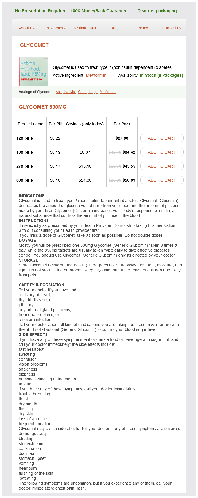
Glycomet 500mg
- 120 pills - $27.00
- 180 pills - $34.42
- 270 pills - $45.55
- 360 pills - $56.69
Glycomet dosages: 500 mg
Glycomet packs: 120 pills, 180 pills, 270 pills, 360 pills
Only $0.17 per item
In stock: 741
Description
The posteriorly placed occipital lobes are separated from the parietal and temporal lobes by the plane of the parieto-occipital sulcus diabetes mellitus type 2 wound healing glycomet 500 mg buy without a prescription, visible on the medial surface of the cerebrum in a hemisected brain. The anteriormost points of the anteriorly projecting frontal and temporal lobes are the frontal and temporal poles. The posteriormost point of the posteriorly projecting occipital lobe is the occipital pole. The frontal lobes occupy the anterior cranial fossae, the temporal lobes occupy the lateral parts of the middle cranial fossae, and the occipital lobes extend posteriorly over the tentorium cerebelli. The diencephalon is composed of the epithalamus, thalamus, and hypothalamus and forms the central core of the brain. The midbrain, the rostral part of the brainstem, lies at the junction of the middle and posterior cranial fossae. The pons is the part of the brainstem between the midbrain rostrally and the medulla oblongata caudally. The medulla oblongata (medulla) is the most caudal subdivision of the brainstem that is continuous with the spinal cord. The cerebellum is the large brain mass lying posterior to the pons and medulla and inferior to the posterior part of the cerebrum. It consists of two lateral hemispheres that are united by a narrow middle part, the vermis. Ventricular System of Brain the ventricular system of the brain consists of two lateral ventricles and the midline 3rd and 4th ventricles connected by the cerebral aqueduct. Each lateral ventricle opens through an interventricular foramen into the 3rd ventricle. The 3rd ventricle, a slit-like cavity between the right and the left halves of the diencephalon. The pyramid-shaped 4th ventricle in the posterior part of the pons and medulla extends inferoposteriorly. Inferiorly, it tapers to a narrow channel that continues into the cervical region of the spinal cord as the central canal. At certain areas on the base of the brain, the arachnoid and pia are widely separated by subarachnoid cisterns. Pontocerebellar cistern (pontine cistern): an extensive space ventral to the pons, continuous inferiorly with the spinal subarachnoid space. Interpeduncular cistern (basal cistern): located in the interpeduncular fossa between the cerebral peduncles of the midbrain. Chiasmatic cistern (cistern of optic chiasma): inferior and anterior to the optic chiasm, the point of crossing or decussation of optic nerve fibers. Quadrigeminal cistern (cistern of great cerebral vein): located between the posterior part of the corpus callosum and the superior surface of the cerebellum; contains parts of the great cerebral vein. Cisterna ambiens (ambient cistern): located on the lateral aspect of the midbrain and continuous posteriorly with the quadrigeminal cistern (not illustrated).
Syndromes
- Recently placed artificial joints
- Sore throat
- Tube through the mouth into the stomach to wash out the stomach (gastric lavage)
- Ovarian cancer
- Heavy bleeding
- Bacterial, viral, or fungal lung infection
- X-rays to determine bone age
- Use of certain medications
- Tuberculosis or other infections
- Yellow eyes
The surface anatomy of the femoral triangle is clinically important because of its contents blood glucose drops rapidly cheap 500 mg glycomet visa. The femoral artery runs a 5-cm superficial course through the femoral triangle before it is covered by the sartorius in the adductor canal. The great saphenous vein enters the thigh posterior to the medial femoral 1638 condyle and passes superiorly along a line from the adductor tubercle to the saphenous opening. The central point of this opening, where the great saphenous vein enters the femoral vein, is located 3. This is one of the most common injuries to the hip region, usually occurring in association with collision sports, such as the various forms of football, ice hockey, and volleyball. Contusions cause bleeding from ruptured capillaries and infiltration of blood into the muscles, tendons, and other soft tissues. The term hip pointer may also refer to avulsion of bony muscle attachments, for example, of the sartorius or rectus femoris to the anterior superior and inferior iliac spines, respectively, of the hamstrings from the ischium. Another term commonly used is "charley horse," which may refer either to the cramping of an individual thigh muscle because of ischemia or to contusion and rupture of blood vessels sufficient enough to form a hematoma. The injury is usually the consequence of tearing of fibers of the rectus femoris; sometimes, the quadriceps tendon is also partially torn. A charley horse is associated with localized pain and/or muscle stiffness and commonly follows direct trauma. The medial arcuate ligament of the diaphragm arches obliquely over the proximal part of the psoas major. The transversalis fascia on the internal abdominal wall is continuous with the psoas fascia, where it forms a fascial covering for the psoas major that accompanies the muscle into the anterior region of the thigh. When the abscess passes between the psoas and its fascia to the inguinal and proximal thigh regions, severe pain may be referred to the hip, thigh, or knee joint. A psoas abscess should always be considered when edema occurs in the proximal part of the thigh. Such an abscess may be palpated or observed in the inguinal region, just inferior or superior to the inguinal ligament, and may be mistaken for an indirect inguinal hernia or a femoral hernia, an enlargement of the inguinal lymph nodes, or a saphenous varix. The lateral border of the psoas is commonly visible in radiographs of the abdomen; an obscured psoas shadow may be an indication of abdominal pathology. Paralysis of Quadriceps A person with paralyzed quadriceps muscles cannot extend the leg against resistance. They commonly walk with a forward lean, pressing on the distal end of the thigh with their hand as the heel contacts the ground to prevent inadvertent flexion of the knee joint. Weakness of the vastus medialis or vastus lateralis, resulting from arthritis or trauma to the knee joint, can result in abnormal patellar movement and loss of joint stability. Such overstressing of the knee region can also occur in running sports such as basketball.
Specifications/Details
The head consists of the brain and its protective coverings (cranial vault and meninges) blood sugar not going up cheap 500 mg glycomet otc, the ears, and the face. The face includes openings and passageways, with lubricating glands and valves (seals) to close some of them, the masticatory (chewing) devices, and the orbits that house the visual apparatus. Disease, malformation, and trauma of structures in the head form the bases of many specialties, including dentistry, maxillofacial surgery, neurology, neuroradiology, neurosurgery, ophthalmology, oral surgery, otology, rhinology, and psychiatry. The neurocranium is the bony case of the brain and its membranous coverings, the cranial meninges. It also contains proximal parts of the cranial nerves and the vasculature of the brain. The neurocranium in adults is formed by a series of eight bones: four singular bones centered on the midline 1871 (frontal, ethmoidal, sphenoidal, and occipital), and two sets of bones occurring as bilateral pairs (temporal and parietal). It may mean the cranium (which includes the mandible) or the part of the cranium excluding the mandible. There has also been confusion because some people have used the term cranium for only the neurocranium. In the anatomical position, the inferior margin of the orbit and the superior margin of the external acoustic meatus lie in the same horizontal orbitomeatal (Frankfort horizontal) plane. The neurocranium and viscerocranium are the two primary functional parts of the 1873 cranium. From the lateral aspect, it is apparent that the volume of the neurocranium, housing the brain, is approximately double that of the viscerocranium. The spinal cord is continuous with the brain through the foramen magnum, the large opening in the basal part of the occipital bone. The viscerocranium, housing the optical apparatus, nasal cavity, paranasal sinuses, and oral cavity, dominates the facial aspect of the cranium. The mandible is a major component of the viscerocranium, articulating with the remainder of the cranium via the temporomandibular joint. The broad ramus and coronoid process of the mandible provide attachment for powerful muscles capable of generating great force in relationship to biting and chewing (mastication). The supra-orbital notch, the infraorbital foramen, and the mental foramen, giving passage to major sensory nerves of the face, are approximately in a vertical line. The neurocranium has a dome-like roof, the calvaria (skullcap), and a floor 1875 or cranial base (basicranium). The bones forming the calvaria are primarily flat bones (frontal, parietal, and occipital; see. The bones contributing to the cranial base are primarily irregular bones with substantial flat portions (sphenoidal and temporal) formed by endochondral ossification of cartilage (chondrocranium) or from more than one type of ossification.
Coing (Quince). Glycomet.
- Are there safety concerns?
- Dosing considerations for Quince.
- How does Quince work?
- What is Quince?
- Are there any interactions with medications?
- Digestive disorders, diarrhea, coughs, stomach and intestinal inflammation, skin injuries, inflammation of the joints, eye discomfort, and other conditions.
Source: http://www.rxlist.com/script/main/art.asp?articlekey=96398
Related Products
Usage: q._h.
Additional information:
Tags: cheap glycomet 500 mg overnight delivery, discount 500 mg glycomet with mastercard, glycomet 500 mg buy with visa, cheap 500 mg glycomet free shipping
9 of 10
Votes: 112 votes
Total customer reviews: 112
Customer Reviews
Wenzel, 46 years: It is a marked thickening of the circular layer of smooth muscle that controls discharge of the stomach contents through the pyloric orifice (inferior opening or outlet of the stomach) into the duodenum. Target organs in decompression sickness include the spinal cord,162 as well as the skin, bone, retina807 and ear.
Givess, 59 years: The anterior border of the left lung has a deep cardiac notch, an indentation consequent to the deviation of the apex of the heart to the left side. The blood vessels of the heart are affected by both sympathetic and parasympathetic nerves.
Asam, 54 years: This plexus gives rise to the five terminal branches of the facial nerve: temporal, zygomatic, buccal, marginal mandibular, and cervical. The obturator artery helps the profunda femoris artery supply the adductor muscles via anterior and posterior branches, which anastomose.
Einar, 33 years: The person is asked if one side feels the same as or different from the other side. However, because of overlapping areas of innervation between nerves, one or two small branches of nerves may usually be cut without a noticeable loss of motor supply to the muscles or loss of sensation to the skin.
Gamal, 55 years: Thus, most muscles are composed of more than one myotome, and most often, multiple spinal cord segments are involved in producing the movement of the lower limb. The thigh muscles are separated into three compartments-anterior, medial, and posterior.