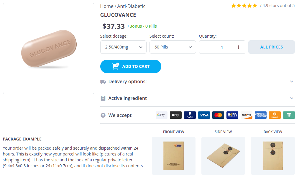
- Glucovance 5/500mg × 60 Pills - $37.33
- Glucovance 5/500mg × 120 Pills - $61.34
- Glucovance 5/500mg × 240 Pills - $109.33
- Glucovance 5/500mg × 300 Pills - $133.34
- Glucovance 2.50/400mg × 60 Pills - $37.33
- Glucovance 2.5/400mg × 120 Pills - $61.34
- Glucovance 2.5/400mg × 240 Pills - $109.33
- Glucovance 2.5/400mg × 300 Pills - $133.34
Glucovance dosages: 5/500 mg, 2.50/400 mg, 2.5/400 mg
Glucovance packs: 60 pills, 120 pills, 240 pills, 300 pills
Only $0.44 per item
In stock: 600
Description
Tibiotalocalcaneal fusion is indicated in patients with arthritis in both the tibiotalar and subtalar joints symptoms appendicitis . The goal of surgical intervention is to obtain a stable, plantigrade, and pain-free foot and ankle. These include (1) to remove medial bony prominences and debris and (2) to assist in resection of medial bone when advanced varus deformity precludes reduction of the foot to neutral. Enter the ankle joint sharply and fully expose it by releasing the lateral ligaments and anterior and posterior capsule. After removing the cartilage, prepare the joint surface with flexible chisels or a small, low-speed burr. Burr holes should be just through the subchondral bone and separated by about 3 mm on all sides to avoid weakening or fracture of the cortex. Curette the remaining cartilage off the joint surface and prepare the subchondral bone with flexible chisels or a burr as described above. If there is significant bone loss or fragmentation of the talus, the tibia may have to be fused directly to the talus. In this case, the calcaneal articular processes will need to be removed with an osteotome to create a flat surface that will lie flush with the tibial plafond. If there is a large bony deficit with substantial loss of limb length, structural graft in the form of iliac crest autograft or femoral head allograft can be used to restore height. The use of both an adolescent blade plate and a humeral blade plate has been described. Ensure that the hindfoot is positioned in neutral to 5 degrees of valgus and the ankle is in neutral dorsiflexion and plantarflexion. The ankle and subtalar joints must be held rigidly during insertion of the blade plate. The guidewire should be inserted such that 5 to 10 mm of calcaneal bone will remain plantar to the blade. The lateral calcaneal cortex may then be further prepared for blade insertion by predrilling with a 4. Contour the plate to the lateral aspect of the tibia and fill the screw holes sequentially. Place the screw under fluoroscopic guidance from the calcaneal tuberosity into the anterior tibial cortex at roughly a 60-degree angle. Further steps that will aid in the prevention of a postoperative hematoma include releasing the tourniquet and assessing hemostasis before closure, the use of drains, and the use of a compression dressing. Once the blade engages the calcaneus, the position of the plate proximally cannot be changed.
Syndromes
- Hyperthyroidism
- Pain that is worse when you lie down or that wakes you up at night
- Those who have not received the vaccine in the past may still get the series of three shots. Talk to your health care provider.
- Bleeding inside the body
- Irritation
- Primary hemochromatosis is a genetic disorder passed down through families. It occurs at birth. People with this condition absorb too much iron through their digestive tract. Iron builds up in the body, especially the liver. You are more likely to get this disease if someone else in your family has or had the condition.
- Severe abdominal pain
- Vertigo that occurs with a fever of more than 101 degrees Fahrenheit
- You develop scars as your acne clears up.
- Jaundice
The guide pin is inserted immediately lateral to the Achilles tendon medicine 1800s , approximately 3 cm proximal to the ankle joint. The second pin is inserted from the anteromedial aspect of the tibia directly above the medial malleolus distally and anteriorly toward the sinus tarsi. The third guide pin is inserted from the lateral aspect of the joint anterior to the fibula and directed toward the medial talar neck. The guide pin is inserted immediately adjacent to the Achilles tendon, approximately 3 cm proximal to the ankle joint. The positions of the first two guide pins are checked under fluoroscopy, and retrieval to appropriate length is performed when necessary. The positions of the guide pins and satisfactory tibiotalar apposition are then checked under fluoroscopy, and appropriate length 6. If there is residual motion in the arthrodesis, I retighten or reposition the screws. With satisfactory stability, I place bone graft at the anterior tibiotalar arthrodesis. After closure of the retinaculum, the residual capsule, subcutanenous tissue, and skin are closed in routine fashion. The screw inserted from the posterolateral tibia is critical because it obtains the best purchase in the talus and is in the plane of the most direct line of compression across the joint. Because the screws are not introduced parallel to each other, eccentric loading of the arthrodesis site may occur as the first one is inserted. This can be avoided by alternately tightening each screw until compression is obtained. Position of the arthrodesis site Preparation of the articular surfaces I aim for neutral position in the sagittal plane, minimal valgus (up to 5 degrees), and external rotation symmetric with the contralateral physiologically normal ankle (no more than 5 to 10 degrees). A high-speed burr and smooth K-wires tend to create localized osteonecrosis that may delay healing. Moreover, the slurry created with a burr may predispose to symptomatic anterior joint synovitis. The joint must be irrigated frequently to visualize the cancellous bone surfaces and confirm uniform bleeding. It is important to position the lamina spreader correctly to avoid tilting the talus from the neutral position. Also, if a midfoot or forefoot deformity compromises optimal positioning of the ankle arthrodesis, adjunctive osteotomies of the foot may be warranted. Alternatively, if a slight modification of the position for ankle arthrodesis may accommodate an associated fixed midfoot or forefoot deformity, such as equinus, it may be preferable to position the ankle in slight dorsiflexion to allow for a plantigrade foot position. Pre-existing hardware Joint preparation When using the miniarthrotomy technique, leave pre-existing hardware in place unless it interferes with insertion of the arthrodesis screws. The miniarthrotomy technique does not afford access to the posterior 25% of the tibiotalar Preparation of only the anterior 75% of the joint leads to fusion rates equal to those of other techniques of ankle arthrodesis in my experience and that of others.
Specifications/Details
The external tibial alignment guide directs the initial tibial cut into 3 degrees of posterior slope; we aim to eliminate this slope symptoms 28 weeks pregnant . To further promote a perpendicular tibial preparation relative to the tibial shaft axis, we raise the proximal aspect of the external tibial alignment guide two to three fingerbreadths above the tibial spine before securing it to the proximal pin. Set the rotation of the cutting block for tibial preparation based on the reference osteotome set in the medial gutter. A dedicated T-guide temporarily attached to the distal aspect of the guide facilitates setting proper rotation. Lock the rotation of the distal block with the knob connecting the telescoping rods of the guide. While controlling rotation, set the proper length of the guide via the telescoping rods. Positioning the proximal pin relative to a reference osteotome placed in the medial gutter. Setting rotation of the distal cutting block of the guide relative to the medial gutter reference osteotome. Angel wing about to be inserted into capture guide attached to distal tibial cutting block. If the initial position of the distal block is set at the apex of the plafond, the desired 5 mm of resection may be easily set and even greater resection is possible in a tighter ankle. We recommend using pins at different levels rather than pins in a single plane (risks creating a stress riser). Attach the cutting capture guide to the distal block, and insert an angel-wing resection guide in the capture guide. Adjust the cutting guide in the coronal plane to ensure that the malleoli are protected with tibial resection. We routinely set the guide based on a pin placed loosely in the medial aspect of the capture guide. We aim to position the guide so that the medial extent of tibial preparation is directly proximal from the transition of tibial plafond to medial malleolus. Drive the pin used as a reference into the tibia through the medial aspect of the capture guide to protect the medial malleolus. Similarly, place a lateral pin in the lateral aspect of the capture guide and advance it into the lateral gutter. The capture guide has several options to place the lateral pin to accommodate any coronal plane dimension of the tibial plafond. With the soft tissues protected, particularly the deep neurovascular bundle, make the distal tibial cut with an oscillating saw through the horizontal portion of the capture guide. A toothless lamina spreader may be placed judiciously on the prepared tibial surface and dorsal talus to facilitate evacuation of bone from the posterior ankle.
Nicotylamidum (Niacin And Niacinamide (Vitamin B3)). Glucovance.
- What other names is Niacin And Niacinamide (vitamin B3) known by?
- Prevention of cataracts, an eye condition.
- Osteoarthritis.
- Treatment and prevention of niacin deficiency, and certain conditions related to niacin deficiency such as pellagra.
- High cholesterol. Only niacin seems to lower cholesterol, not niacinamide.
- Dosing considerations for Niacin And Niacinamide (vitamin B3).
Source: http://www.rxlist.com/script/main/art.asp?articlekey=96888
Related Products
Usage: q.h.
Additional information:
Tags:
9 of 10
Votes: 216 votes
Total customer reviews: 216
Customer Reviews
Raid, 60 years: Avoiding wound complications after neglected Achilles tendon repair: tissue expansion technique. After attaching the graft to the distal aspect of the capsule, perform the standard Brostrom repair. We therefore recommend that the mini-open technique be performed through a short longitudinal incision that can easily be extended if necessary.
Kirk, 63 years: The more conservative arthrodesis that maintains subchondral bone architecture of the joints is reserved for mild to moderate deformity. The inferior half of the posterior calcaneal tuberosity has a rough surface with an extensive Sharpey fiber network. Active plantarflexion of the foot is usually preserved due to the action of tibialis posterior, the peroneal tendons, and the long toe flexors.
Onatas, 50 years: The origin of the extensor digitorum brevis muscle is identified and elevated along with the sinus tarsi fat pad as a distally based flap. Both endplates should be thoroughly denuded of cartilage and decorticated to reveal bleeding bony surfaces to enhance the chance of successful fusion. The tip of the superior articular process and the lateral pars interarticularis are exposed with electrocautery.
Farmon, 23 years: The navicular tunnel is made as large as possible without fracturing the navicular to enable placement of large grafts. Active infection or osteomyelitis is a contraindication for this technique as the hardware is typically permanent and difficult or impossible to remove without significant bony destruction. As in all lumbar spinal surgical procedures, if trouble is encountered locating a nerve root, the surgeon should find or palpate the associated pedicle and look along the medial and inferior pedicle wall.
Arakos, 28 years: The nerve injury can be difficult to discern and may be associated with other pathology, which is usually addressed at the same surgical procedure. Degenerative spondylolisthesis should be treated by spinal fusion with or without instrumentation, as discussed in following chapters. Since the extremity will tend to be slightly shorter than the contralateral leg, aligning the second ray of the foot with the anterior tibial crest is appropriate and allows for adequate clearance with a heel-to-toe gait.